
 Explore BioBoost Labs
Muscle & Performance
How skeletal muscle grows, repairs, and fuels itself — and what the research literature actually documents about the compounds studied in this space.
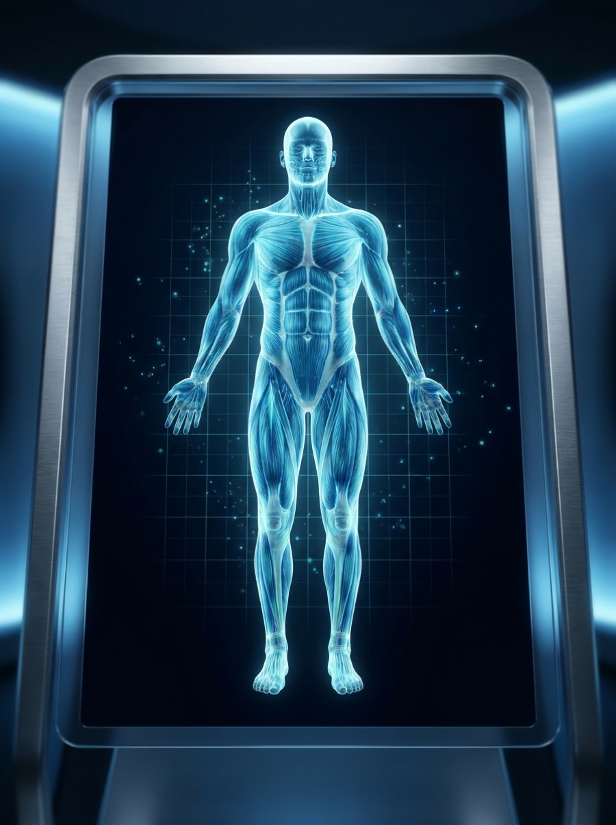
The system at a glance
Skeletal muscle is built from long multinucleated fibers that thicken (hypertrophy) or shrink (atrophy) depending on the balance between protein synthesis and breakdown. Growth signals switch on the PI3K/Akt/mTOR pathway to make new muscle protein, while resident muscle stem cells (satellite cells) supply fresh nuclei needed for repair and enlargement. Overlaying this is the growth-hormone (GH) axis and the mitochondria that generate the energy for contraction and exercise adaptation. Most compounds grouped here act on one of these levers: the GH/IGF-1 signal, satellite-cell activity, or mitochondrial and fat-energy metabolism.
How it signals
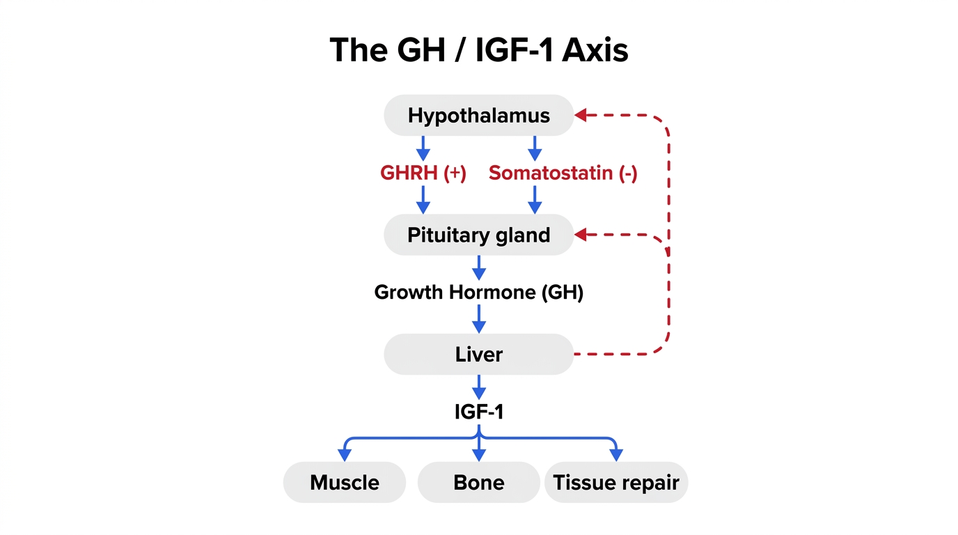
The key messengers are growth hormone (released in pulses from the pituitary) and insulin-like growth factor 1 (IGF-1), which drives muscle protein synthesis through the PI3K/Akt/mTOR and Akt/GSK3 pathways. Pituitary GH release is controlled by two receptors: the GHRH receptor (targeted by GHRH-analogs such as sermorelin and CJC-1295) and the growth-hormone-secretagogue / ghrelin receptor (targeted by ipamorelin). Satellite cells respond to IGF-1 by proliferating and differentiating, while mitochondrial signaling peptides and NAD-related enzymes (such as NNMT) tune cellular energy, AMPK activity, and fat oxidation.
Research peptides studied in this system
Each card summarizes the documented mechanism, what was actually studied and in what model, and how strong the evidence is. These are descriptions of laboratory research — not recommendations, and not evidence of benefit in humans. According to research indexed in PubMed:
What we don’t know — and the risks
Honest limits matter as much as the mechanisms. For this system specifically:
- Compounds that stimulate the GH axis (CJC-1295, ipamorelin, sermorelin) raise GH and IGF-1; sustained supraphysiologic GH/IGF-1 signaling is linked to insulin resistance, fluid retention, and tissue overgrowth in GH-excess states — not characterized for these peptides at self-administered exposures.
- IGF-1 LR3 resists binding proteins and signals systemically; no controlled human trials define a safe muscle-building exposure, and persistent mitogenic signaling raises theoretical cell-proliferation concerns.
- Most muscle and performance findings here come from rodent, cell-culture, or non-muscle endpoints; extrapolation to human muscle mass, strength, or body composition is unproven.
- The CJC-1295 + ipamorelin combination has not been evaluated in controlled trials for efficacy or long-term safety, and the widely referenced human CJC-1295 data used the long-acting DAC form, not the no-DAC version.
- 5-Amino-1MQ and AOD-9604 muscle/metabolic evidence is essentially preclinical; human effect sizes, dosing, and long-term safety are undefined.
- Research-grade peptide identity, purity, and dose are not guaranteed, and long-term outcomes for all of these compounds remain unstudied.
Responsible understanding
◆ This is education, not medical advice
This page is educational and describes what has been studied in laboratory and clinical research. It is not medical advice, and these materials are for research use only — not for human or veterinary use. Timing, administration, and whether anything is used at all are clinical decisions that belong with a licensed physician overseeing your care; we take no position on them. Science and regulation evolve; verify anything important against the primary sources below.
Sources
Based on articles retrieved from PubMed. Follow each link to the original paper.
- Rommel C et al. Mediation of IGF-1-induced skeletal myotube hypertrophy by PI(3)K/Akt/mTOR and PI(3)K/Akt/GSK3 pathways. Nature Cell Biology. 2001;3(11):1009-13. DOI
- Mouly V et al. The mitotic clock in skeletal muscle regeneration, disease and cell mediated gene therapy. Acta Physiologica Scandinavica. 2005;184(1):3-15. DOI
- Gao B et al. Changes in Skeletal Muscle and Body Weight on Sleeping Beauty Transposon-Mediated Transgenic Mice Overexpressing Pig mIGF-1. Biochemical Genetics. 2018;56(4):341-355. DOI
- Teichman SL et al. Prolonged stimulation of growth hormone (GH) and insulin-like growth factor I secretion by CJC-1295, a long-acting analog of GH-releasing hormone, in healthy adults. The Journal of Clinical Endocrinology and Metabolism. 2006;91(3):799-805. DOI
- Johansen PB et al. Ipamorelin, a new growth-hormone-releasing peptide, induces longitudinal bone growth in rats. Growth Hormone & IGF Research. 1999;9(2):106-13. DOI
- Chen RG et al. A comparative study of growth hormone (GH) and GH-releasing hormone(1-29)-NH2 for stimulation of growth in children with GH deficiency. Acta Paediatrica Supplement. 1993;388:32-5. DOI
- Lee C et al. The mitochondrial-derived peptide MOTS-c promotes metabolic homeostasis and reduces obesity and insulin resistance. Cell Metabolism. 2015;21(3):443-54. DOI
- Reynolds JC et al. MOTS-c is an exercise-induced mitochondrial-encoded regulator of age-dependent physical decline and muscle homeostasis. Nature Communications. 2021;12(1):470. DOI
- Neelakantan H et al. Selective and membrane-permeable small molecule inhibitors of nicotinamide N-methyltransferase reverse high fat diet-induced obesity in mice. Biochemical Pharmacology. 2017;147:141-152. DOI
- Heffernan MA et al. Increase of fat oxidation and weight loss in obese mice caused by chronic treatment with human growth hormone or a modified C-terminal fragment. International Journal of Obesity and Related Metabolic Disorders. 2001;25(10):1442-9. DOI
Research-use-only educational content. Nothing here is medical, dosing, timing, or treatment advice.