
 Explore BioBoost Labs
The Musculoskeletal System
How bone, muscle, tendon and ligament move you — and what the research literature actually reports about the compounds studied in tissue repair here.
The system at a glance
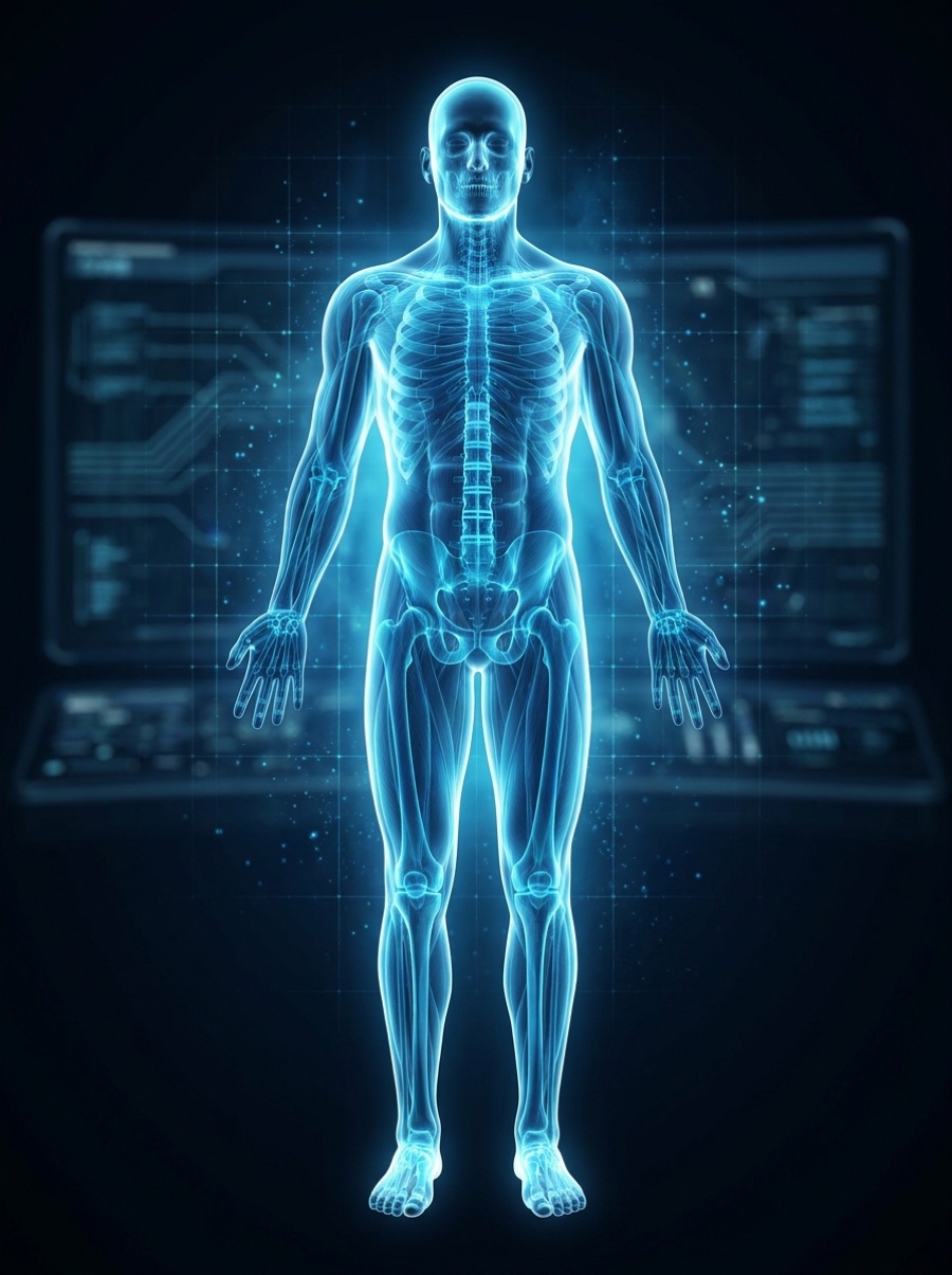
The musculoskeletal system is the body’s framework and its engine. Bones give structure and protect organs; skeletal muscles contract to produce movement; and tendons and ligaments — dense, collagen-rich connective tissue — transmit that force and hold joints together. Because much of this tissue is poorly supplied with blood, it can be slow to recover after injury.
When these tissues are damaged, the body runs a coordinated repair program: an early inflammatory phase clears debris, resident stem cells (in muscle, the satellite cells) proliferate, new blood vessels grow in, and collagen is laid down and then remodeled back toward the original architecture.12 Much of the interest in peptides here centers on that repair cascade.
How it signals
Repair is directed by chemical messengers. A few names recur throughout the compound research below, so they are worth knowing first:
Growth factors — proteins such as IGF-1, VEGF and FGF that tell cells to divide, migrate or build tissue. Angiogenesis — the growth of new blood vessels into a healing site, which many of these compounds are studied to influence. Satellite cells — muscle’s resident stem cells, which wake up after injury to rebuild muscle fibers. The extracellular matrix (ECM) — the collagen scaffold that gives tendon and ligament their strength; healing is largely a story of how well that scaffold is rebuilt.128
Research peptides studied in this system
Each card summarizes the documented mechanism, what was actually studied and in what model, and how strong the evidence is. These are descriptions of laboratory research — not recommendations, and not evidence of benefit in humans. According to research indexed in PubMed:
What we don’t know — and the risks
Honest limits matter as much as the mechanisms. For this system specifically:
- Most evidence is preclinical. The strongest musculoskeletal results (especially for BPC-157) come from rodent models. Animal healing does not reliably predict human outcomes.3
- Human data is borrowed from other tissues. TB-500 and GHK-Cu human trials studied skin, cornea and heart — not tendon, ligament or muscle. Applying those results to musculoskeletal use is an assumption, not a finding.56
- Growth signaling cuts both ways. IGF-1 pathways that build muscle also influence cell proliferation broadly; potent growth-factor signaling carries theoretical risks that have not been characterized for these research analogs.8
- Purity and identity are unknowns. Research-grade material is not a medicine. Without independent testing, what is actually in a vial — identity, purity, contamination, endotoxin — is not guaranteed.
- Long-term effects are unstudied. There is essentially no long-term human safety data for these compounds in a musculoskeletal context.
- Individual variability and interactions. Response and risk vary between individuals, and interactions with medications or conditions are not mapped.
Responsible understanding
◆ This is education, not medical advice
This page is educational and describes what has been studied in laboratory and clinical research. It is not medical advice, and these materials are for research use only — not for human or veterinary use. Anyone considering anything related to their own health should consult a licensed physician who can weigh their individual history and risks. Science and regulation evolve; verify anything important against the primary sources below.
Sources
Based on articles retrieved from PubMed. Follow each DOI link to the original paper.
- Tidball JG. Mechanisms of muscle injury, repair, and regeneration. Compr Physiol. 2011;1(4):2029–62. DOI
- Yoshimoto Y, Oishi Y. Mechanisms of skeletal muscle-tendon development and regeneration/healing as potential therapeutic targets. Pharmacol Ther. 2023;243:108357. DOI
- Gwyer D, Wragg NM, Wilson SL. Gastric pentadecapeptide body protection compound BPC 157 and its role in accelerating musculoskeletal soft tissue healing. Cell Tissue Res. 2019;377(2):153–159. DOI
- Seiwerth S, et al. BPC 157 and Standard Angiogenic Growth Factors… Tendon, Ligament, Muscle and Bone Healing. Curr Pharm Des. 2018;24(18):1972–1989. DOI
- Goldstein AL, Hannappel E, Sosne G, Kleinman HK. Thymosin β4: a multi-functional regenerative peptide. Basic properties and clinical applications. Expert Opin Biol Ther. 2012;12(1):37–51. DOI
- Pickart L. The human tri-peptide GHK and tissue remodeling. J Biomater Sci Polym Ed. 2008;19(8):969–88. DOI
- Pickart L, Vasquez-Soltero JM, Margolina A. The human tripeptide GHK-Cu in prevention of oxidative stress and degenerative conditions of aging. Oxid Med Cell Longev. 2012;2012:324832. DOI
- Yoshida T, Delafontaine P. Mechanisms of IGF-1-Mediated Regulation of Skeletal Muscle Hypertrophy and Atrophy. Cells. 2020;9(9):1970. DOI
- Musarò A, et al. Localized Igf-1 transgene expression sustains hypertrophy and regeneration in senescent skeletal muscle. Nat Genet. 2001;27(2):195–200. DOI
Research-use-only educational content. Nothing here is medical, dosing, or treatment advice.