

Cellular Health & Longevity

Body & Peptide Science · System 08

Cellular Health & Longevity

How the literature describes telomere biology, cellular senescence, mitochondrial-derived peptides, and redox balance — mechanism only, most of it preclinical.
5 compounds studied9 primary sourcesResearch use only
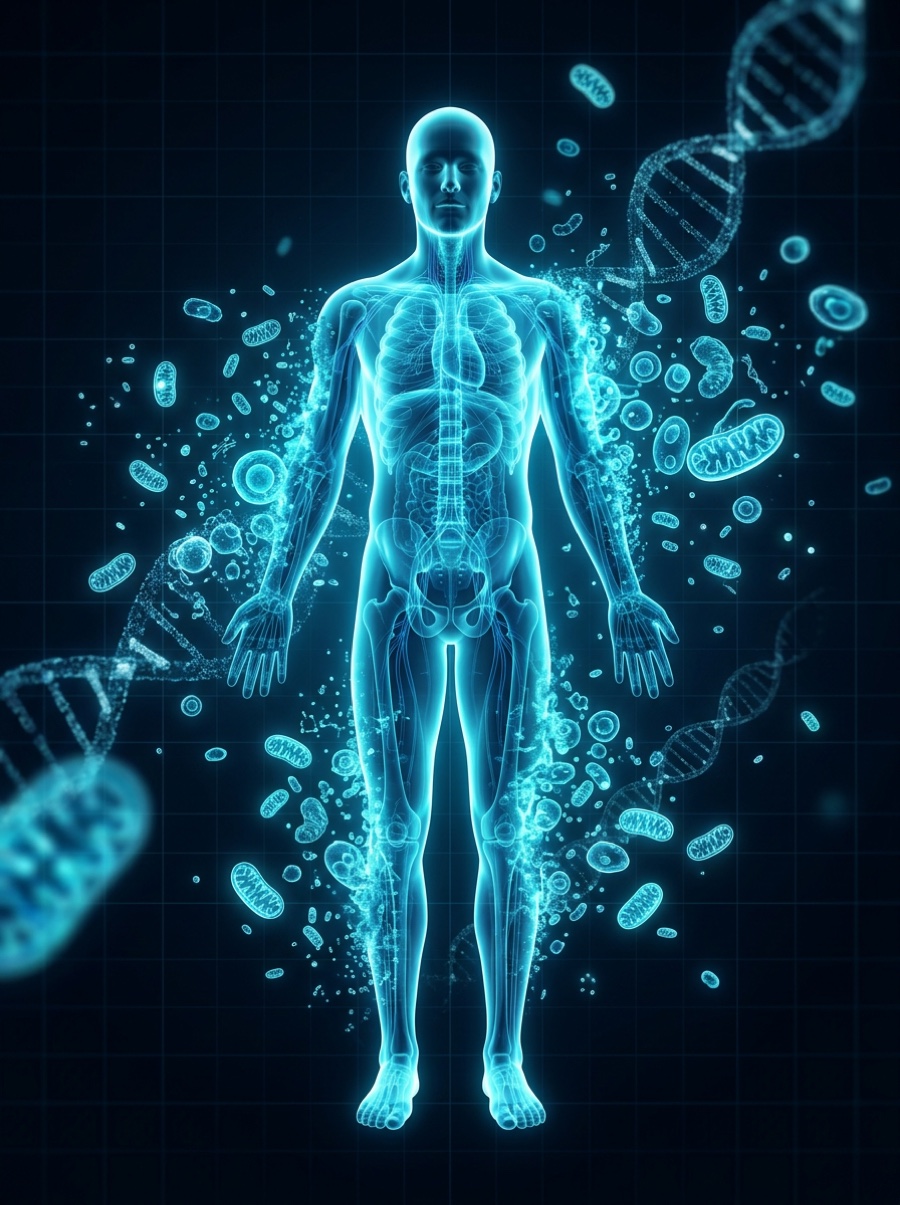
Anatomical bio-scan of the Cellular Health & Longevity
01

The system at a glance

“Aging” is not one process but several overlapping ones happening inside cells: chromosome-end (telomere) shortening that limits how many times a cell can divide; the buildup of “senescent” cells that stop dividing but linger and secrete inflammatory signals; a decline in the mitochondrial and metabolic cofactors that power the cell; and a drift in redox balance, the tug-of-war between reactive oxygen species and antioxidant defenses. The compounds on this page are studied as tools to probe those mechanisms — a telomerase-associated peptide (Epithalon), an engineered senescent-cell-killing peptide (FOXO4-DRI), a redox and signaling coenzyme (NAD+), a master cellular antioxidant (Glutathione), and a mitochondria-encoded metabolic peptide (MOTS-c). This is frontier science. Almost all of the longevity-relevant evidence comes from cells in a dish and from mice, and the reader should treat every mechanism below as a research hypothesis, not an established human effect.

02

How it signals

This is one of the most hype-prone corners of biology, so calibrate hard against the evidence. There are NO human trials showing any of these compounds extend human lifespan or reverse human aging — none exist, and any claim otherwise is unsupported. A mechanism demonstrated in cultured cells or in mice frequently fails to reproduce in people. Several of these agents (Epithalon, MOTS-c, FOXO4-DRI) rest on a small, often single-lab or preclinical evidence base. Effects seen at controlled doses in animals say nothing about safety or benefit in humans. Everything here is provided strictly for research and educational context under research-use-only conditions; none of it is a therapy, a supplement recommendation, or medical advice.

03

Research peptides studied in this system

Each card summarizes the documented mechanism, what was actually studied and in what model, and how strong the evidence is. These are descriptions of laboratory research — not recommendations, and not evidence of benefit in humans. According to research indexed in PubMed:

EpithalonEpitalon; Ala-Glu-Asp-Gly (AEDG tetrapeptide)Low — small, largely single-lab preclinical body
Documented mechanism
A synthetic four-amino-acid peptide reported to interact with the telomerase promoter region and induce expression of the telomerase catalytic subunit (hTERT), the enzyme that rebuilds telomeres — the protective DNA caps that shorten each time a cell divides. The proposed idea is that restoring telomerase activity lengthens telomeres and delays replicative senescence (the “Hayflick limit”). This is a mechanistic hypothesis, not a demonstrated human outcome. 1
What was actually studied
Cultured normal human diploid fibroblasts, where treated cells reportedly elongated telomeres and completed additional cell divisions beyond untreated controls; plus proposed molecular models of peptide-DNA binding at the telomerase promoter. The evidence base is small and comes largely from a single research group.
Evidence tier
Telomerase re-activation is a double-edged concept: the same enzyme that could extend cellular lifespan is also exploited by most cancers, which is a core safety question raised in the field. No human study demonstrates lifespan extension or anti-aging benefit. Treat all claims here as unproven.
FOXO4-DRIFOXO4 D-retro-inverso peptide; proxofimLow — proof-of-concept preclinical (cells + mice)
Documented mechanism
An engineered cell-penetrating peptide designed as a ‘senolytic’ — a molecule that selectively kills senescent cells. It disrupts the interaction between the transcription factor FOXO4 and the tumor-suppressor protein p53. In senescent cells, this releases p53 from the nucleus and triggers their self-destruction (apoptosis), while sparing healthy dividing cells. The goal studied is to clear the lingering senescent cells thought to drive tissue aging and chronic inflammation. 23
What was actually studied
The founding work was in cultured cells and in mice — including fast-aging (Xpd) and naturally aged mice, where the peptide was reported to restore fur density, fitness, and kidney function, and to blunt chemotherapy toxicity. More recent cell and tissue-culture work has examined it in senescent human fibroblasts and keloid scar models. All in vitro or animal; no human efficacy data.
Evidence tier
This is a designed research peptide, not an approved drug, and the senolytic field broadly is still early. Selectively removing senescent cells has plausible upside and real unknowns (senescent cells also play roles in wound healing and tumor suppression). No human lifespan or anti-aging evidence exists.
NAD+Nicotinamide adenine dinucleotide (oxidized form)Moderate — deep mechanistic base; human longevity outcomes unproven
Documented mechanism
A central coenzyme in every cell. In its redox role it shuttles electrons through energy metabolism (glycolysis, the TCA cycle, oxidative phosphorylation). Separately, it is the required fuel for a set of ‘consumer’ enzymes — sirtuins, PARPs (DNA repair), and CD38 — that regulate DNA repair, chromatin, gene expression, and cellular senescence. Tissue NAD+ levels are documented to decline with age across model organisms, and that decline is hypothesized to contribute to multiple aging-associated dysfunctions. 45
What was actually studied
An extensive mechanistic literature in cells and rodents links NAD+ decline to mitochondrial dysfunction, impaired DNA repair, and metabolic disease, and shows that restoring NAD+ (often via precursors) can improve age-related functional measures in animals. Human data exist mostly for NAD+ precursors and focus on biomarkers and specific conditions — not on lifespan.
Evidence tier
The biochemistry is well established; the leap that is NOT established is that raising NAD+ slows human aging or extends human lifespan. Reviewers in the field explicitly flag open questions about whether repletion is beneficial and safe in aging humans.
GlutathioneGSH; L-γ-glutamyl-L-cysteinylglycineModerate — established redox biology; anti-aging claims unproven
Documented mechanism
The cell’s most abundant small-molecule antioxidant, a tripeptide (glutamate-cysteine-glycine). It neutralizes reactive oxygen and nitrogen species, recycles other antioxidants, and cycles between reduced (GSH) and oxidized (GSSG) forms; that ratio is a core readout of a cell’s ‘redox balance.’ The aging-relevant hypothesis is that a drift toward oxidative stress — too many reactive species relative to antioxidant capacity — damages lipids, proteins, and DNA and helps drive senescence and inflammation. 67
What was actually studied
Redox biology and glutathione’s role are well characterized across cell and animal studies and in human disease contexts. Clinical antioxidant work has largely used glutathione-generating agents such as N-acetylcysteine (e.g., in COPD) rather than glutathione’s aging role directly; dietary/antioxidant interventions have a mixed and often disappointing clinical track record.
Evidence tier
Oxidative stress is real biology, but the simple ‘more antioxidants = slower aging’ story has repeatedly failed to hold up in humans, and reactive oxygen species also serve necessary signaling functions. No evidence supports a human anti-aging or lifespan effect.
MOTS-cMitochondrial ORF of the 12S rRNA type-c (mitochondrial-derived peptide)Low — early preclinical (cells + mice)
Documented mechanism
A short peptide encoded within mitochondrial DNA — one of the first examples of the mitochondrial genome directly signaling to the rest of the cell. It is reported to activate AMPK (a master energy-sensing kinase), influence the folate/purine metabolic cycle, and, under metabolic stress, translocate to the nucleus where it interacts with stress-responsive transcription factors including NRF2 to regulate antioxidant-response genes. It is studied as a signal that helps coordinate metabolism between mitochondria and the nucleus. 89
What was actually studied
Cultured cells and mice: in mice it was reported to improve insulin sensitivity and resist high-fat-diet-induced obesity and age-dependent insulin resistance; other rodent work examines nuclear signaling, antioxidant-gene regulation, and nerve-injury models. All preclinical — no human efficacy or longevity data.
Evidence tier
MOTS-c is a genuinely interesting discovery in mitochondrial biology, but the longevity framing outruns the evidence: the work is early, largely from a small number of labs, and entirely non-human for the outcomes people care about.
04

What we don’t know — and the risks

Honest limits matter as much as the mechanisms. For this system specifically:

  • No human lifespan or anti-aging evidence: not a single one of these compounds has a human trial showing extended lifespan, reversed aging, or a longevity benefit — and for several, no human efficacy data exist at all.
  • Preclinical does not equal proven: most findings here are from cultured cells or mice, and mechanisms that work in those systems very often fail to translate to people.
  • Small or single-lab evidence: Epithalon, MOTS-c, and FOXO4-DRI rest on limited, sometimes single-group bodies of work that have not been broadly independently replicated.
  • Mechanisms cut both ways: telomerase re-activation intersects with cancer biology, senolytics can hit cells with useful functions, and antioxidants can blunt necessary signaling — ‘more’ is not automatically ‘better.’
  • Purity, identity, and dosing are undefined in a research context: these are research-use-only materials, not standardized therapies, and this page gives no dosing, timing, or protocol guidance of any kind.
  • Not medical advice: nothing here is a treatment recommendation or a substitute for evaluation and oversight by a qualified physician.
05

Responsible understanding

◆ This is education, not medical advice

This page is educational and describes what has been studied in laboratory and clinical research. It is not medical advice, and these materials are for research use only — not for human or veterinary use. Timing, administration, and whether anything is used at all are clinical decisions that belong with a licensed physician overseeing your care; we take no position on them. Science and regulation evolve; verify anything important against the primary sources below.

06

Sources

Based on articles retrieved from PubMed. Follow each link to the original paper.

  1. Khavinson VKh, Bondarev IE, Butyugov AA, Smirnova TD Peptide promotes overcoming of the division limit in human somatic cell Bulletin of Experimental Biology and Medicine. 2004;Bull Exp Biol Med. 2004;137(5):503-6. DOI
  2. Baar MP, Brandt RMC, Putavet DA, et al. Targeted Apoptosis of Senescent Cells Restores Tissue Homeostasis in Response to Chemotoxicity and Aging Cell. 2017;Cell. 2017;169(1):132-147.e16. DOI
  3. Kong YX, Li ZS, Liu YB, Pan B, Fu X, Xiao R, Yan L FOXO4-DRI induces keloid senescent fibroblast apoptosis by promoting nuclear exclusion of upregulated p53-serine 15 phosphorylation Communications Biology. 2025;Commun Biol. 2025;8(1):299. DOI
  4. Covarrubias AJ, Perrone R, Grozio A, Verdin E NAD+ metabolism and its roles in cellular processes during ageing Nature Reviews Molecular Cell Biology. 2021;Nat Rev Mol Cell Biol. 2021;22(2):119-141. DOI
  5. Imai S, Guarente L NAD+ and sirtuins in aging and disease Trends in Cell Biology. 2014;Trends Cell Biol. 2014;24(8):464-71. DOI
  6. Valko M, Leibfritz D, Moncol J, Cronin MTD, Mazur M, Telser J Free radicals and antioxidants in normal physiological functions and human disease The International Journal of Biochemistry & Cell Biology. 2007;Int J Biochem Cell Biol. 2007;39(1):44-84. DOI
  7. Barnes PJ Oxidative stress-based therapeutics in COPD Redox Biology. 2020;Redox Biol. 2020;33:101544. DOI
  8. Lee C, Zeng J, Drew BG, et al. The mitochondrial-derived peptide MOTS-c promotes metabolic homeostasis and reduces obesity and insulin resistance Cell Metabolism. 2015;Cell Metab. 2015;21(3):443-54. DOI
  9. Kim KH, Son JM, Benayoun BA, Lee C The Mitochondrial-Encoded Peptide MOTS-c Translocates to the Nucleus to Regulate Nuclear Gene Expression in Response to Metabolic Stress Cell Metabolism. 2018;Cell Metab. 2018;28(3):516-524.e7. DOI
Body & Peptide Science · Cellular Health & Longevity · Draft for review
Research-use-only educational content. Nothing here is medical, dosing, timing, or treatment advice.
For research use only — not for human consumption, and not for diagnosing, treating, curing, or preventing any disease or condition, or making any medical claim. BioBoost Research is an educational resource. Science and regulation are evolving, and the information here may be incomplete, become outdated, or contain errors. Nothing here is medical, legal, or dosing advice — always verify against primary sources and consult a qualified professional. All information reflects facts as best known to BioBoost Research. Full disclaimer →

On clinical data and dosing: Any clinical trials, data, or dosing figures referenced anywhere on this site were conducted in controlled settings under qualified professional and physician oversight, and are shown for informational and educational purposes only — never as guidance. BioBoost Research makes no claim that the same outcome or safety profile would apply to any compound, person, or context. Research and educational use only · 21+.

Read the full Disclaimer →